Mitral valve surgery is performed for two main reasons: the valve has become calcified and narrowed, requiring removal and replacement with a prosthetic valve; or the valve has lost its function due to structural deterioration, resulting in mitral valve regurgitation, which necessitates surgery.
If the valve is severely narrowed and calcified, replacement is necessary. However, if the valve is structurally damaged and causing heart failure, the gold standard procedure is repair rather than replacement. This is because, when the heart valve is repaired, you don’t have to take blood thinners for the rest of your life. Studies in this field show that valve repair is a surgical method that better preserves heart function.
- It is usually caused by rheumatic fever following a severe respiratory infection in childhood that was not properly treated.
- The mitral valve leaflets thicken and become calcified. The leaflets stick together and can no longer move properly.
- The normal mitral valve opening is between 4–6 cm². When the valve opening narrows to less than 1.5 cm², it is considered significant stenosis that requires surgery.
- Because the valve cannot open fully, blood coming from the lungs cannot completely empty into the left ventricle. This leads to increased pressure in the veins coming from the lungs. Fluid builds up in the lungs (pulmonary edema), causing shortness of breath.
- If surgery is delayed, irreversible high pressure in the pulmonary vessels and impaired heart contraction can develop. At this stage, surgery becomes highly risky or may no longer be beneficial (shortness of breath may persist even after valve replacement).
- It can be caused by rheumatic fever, a heart attack, connective tissue diseases, or advanced age.
- Mitral regurgitation may occur when the thread-like structures (like parachute cords) that connect the mitral valve leaflets to the heart muscle rupture, or when the ring (annulus) to which the leaflets are attached dilates, preventing the leaflets from meeting properly.
- When the heart contracts, not all the blood is pumped out to the body; some leaks backward into the lungs through the improperly closing mitral valve. This increases blood pressure in the lungs, leading to shortness of breath.
- In both mitral stenosis and regurgitation, the left atrium enlarges. As a result, heart rhythm disorders, such as atrial fibrillation, may develop. In this irregular rhythm, blood flow inside the heart is disrupted, which can lead to clot formation. If a clot travels to the brain, it can cause a stroke.
- In cases of mitral stenosis, the calcified and immobile valve must be removed. A prosthetic mitral valve is then implanted in its place.
- This prosthetic valve can be made of metal (such as titanium) or it can be a biological valve prepared from bovine tissue made biocompatible for human use.
- Patients with a metal valve must take a blood thinner (Warfarin) for life. In contrast, biological valves do not carry a long-term clotting risk, so anticoagulants are not needed beyond the first three months.
- In mitral regurgitation, the goal is to repair the valve. Since your own valve is preserved, heart function is better maintained over the long term compared to valve replacement. Additionally, since no prosthetic is used, there is no need for long-term anticoagulant therapy.
- There are various valve repair techniques. Your surgeon will determine and explain the best approach for your specific condition.
- If the valve disease is accompanied by an arrhythmia such as atrial fibrillation, a rhythm correction procedure (ablation) should be performed during the same surgery.
- Traditionally, mitral valve surgeries are performed from the front by cutting through the breastbone (sternotomy), using a heart-lung machine.
- In minimally invasive procedures, the mitral valve can be repaired or replaced through a small 4–5 cm incision made under the right breast.
- In endoscopic surgery, the same procedures are performed as in open surgery — just with less trauma to the chest.
Minimally invasive mitral valve surgeries are carried out through a 4–6 cm incision under the right breast and a 2 cm incision in the groin. Both valve repair and replacement (with a prosthetic valve) can be performed through the same incision.
Using special long cannulas inserted through the groin, the heart is connected to a heart-lung machine. The surgery is performed with specially designed instruments and a camera system tailored for these procedures.
With this technique, tricuspid valve repairs, cardiac tumor removals (such as myxomas), and congenital heart defect closures (like atrial septal defects) can also be performed.
- Your doctor should inform you about your condition as if you’re hearing it for the first time, providing complete and clear explanations.
- They should discuss all treatment options with you — both surgical and non-surgical — and their outcomes (medication, monitoring, surgery, valve repair or replacement).
- If surgery is being considered, they should explain the different options and which one they recommend for your specific situation (valve repair or replacement, mechanical vs. biological prosthetic valves, robotic, minimally invasive, or open surgery).
- The doctor should walk you through the entire process starting from the preoperative period, including possible risks.
- Additionally, a dental consultation should be requested to check for any dental issues before surgery.
Typically, patients are admitted to the hospital one day before surgery for a series of routine tests. These tests are conducted to identify any conditions that may pose risks during the operation and to take necessary precautions. They usually include comprehensive blood work, echocardiography, carotid artery ultrasound, chest X-ray or CT scan, and a pulmonary function test.
- If you are taking blood-thinning medications, they generally need to be stopped 5–7 days prior to surgery (Aspirin usually does not need to be discontinued). Other medications will be adjusted by your doctor.
- Body hair removal will be done by hospital staff using specialized tools according to the surgical plan — do not attempt to shave yourself. A nurse will explain how to bathe using a special antiseptic soap.
- The night before the surgery, your doctor may prescribe a sedative to help ensure you get a good night's sleep.
- As you are taken to the operating room, you will remain conscious, but due to a sedative administered through an IV, you won’t remember entering the OR.
- Heart surgeries, including valve procedures, are performed under general anesthesia. The anesthesia process typically takes about 45–50 minutes before the surgery begins.
- Electrodes are placed on your back to monitor your heart rhythm throughout the procedure. A small IV line is inserted into your arm, and a special arterial catheter is placed in your wrist to continuously monitor your blood pressure during surgery and in the intensive care unit.
- Thanks to a sedative injection given before you're taken to the operating room, these procedures are painless and you won’t remember them.
- General anesthesia is administered through intravenous medication. Once full anesthesia is achieved, a tube is inserted into your windpipe (trachea), and your breathing is managed by a ventilator.
- Heart surgeries are performed under general anesthesia, which takes about 45–50 minutes to administer prior to the actual operation.
- Electrodes are placed on your back to monitor your heart rhythm during the surgery. A small intravenous (IV) line is inserted into your arm, and a special arterial catheter is placed in your wrist to continuously monitor your blood pressure during the operation and throughout your stay in the intensive care unit. Thanks to the sedative injection administered before being taken to the operating room, these steps are painless and will not be remembered.
- General anesthesia is induced through IV medications. Once full anesthesia is achieved, a breathing tube is inserted into your trachea, and a ventilator will maintain your breathing throughout the procedure.
- A larger catheter is inserted into a vein in your neck for the administration of medications and fluids during and after the surgery. Blood samples can also be taken through this line, eliminating the need for repeated needle sticks. A urinary catheter is placed as well.
- To evaluate your heart, particularly valve function during and after surgery, a special ultrasound probe is inserted into your esophagus for transesophageal echocardiography.
- Your body is positioned appropriately for the procedure, and the surgical area is disinfected with special antiseptic solutions.
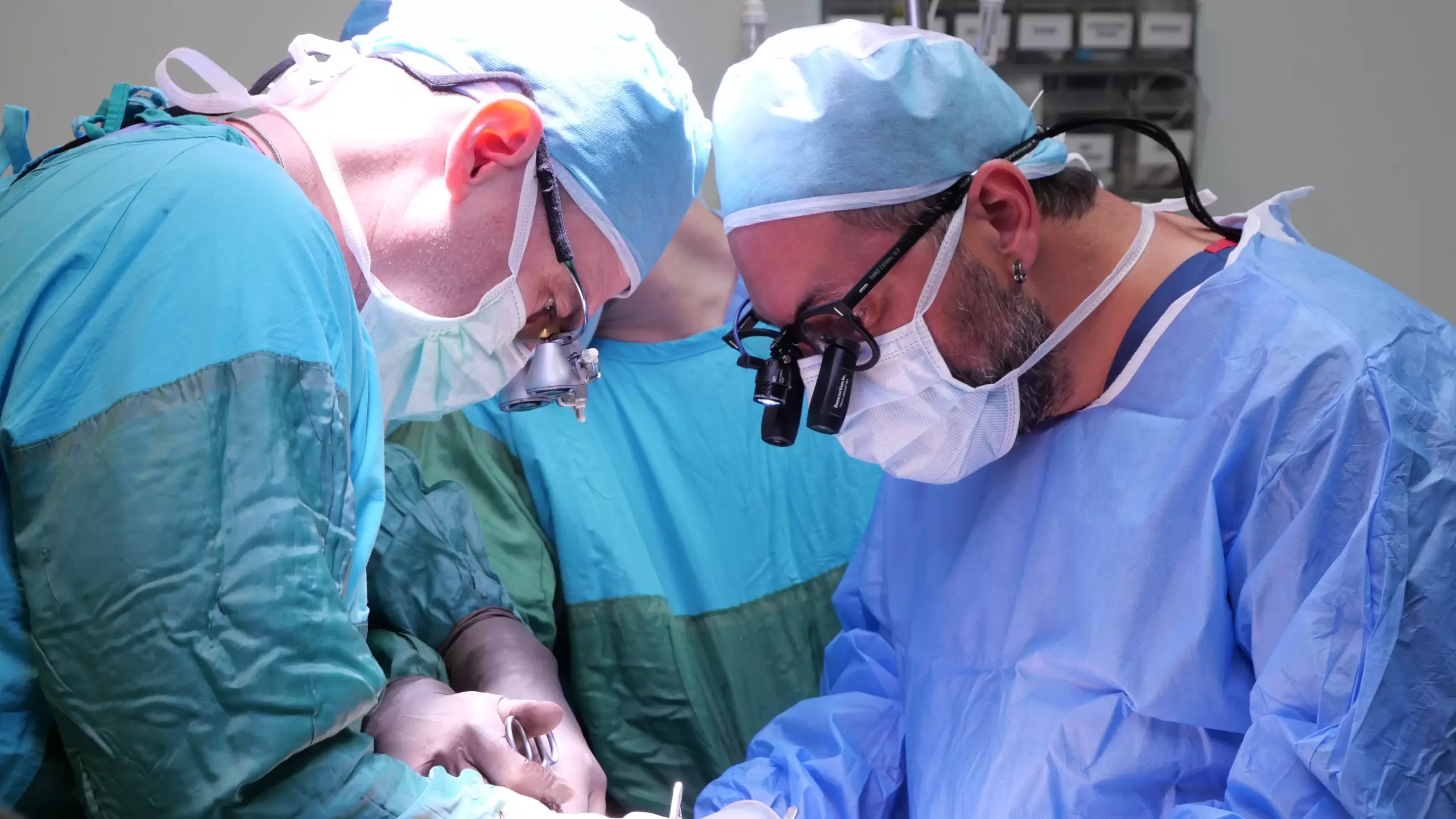
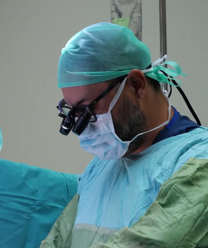
- The surgery then begins… From this moment on, your surgeon is assisted by two other surgeons. One scrub nurse (in sterile attire) and another circulating nurse are present. An anesthesiologist and a technician are continuously at your side. Two perfusionists are responsible for operating the heart-lung machine that temporarily takes over the function of your heart and lungs during the procedure.
- After the surgery is completed, you will be transferred to the intensive care unit while still under anesthesia and connected to a ventilator.
- You will not regain consciousness in the operating room. Depending on your overall condition, you will be gently, safely, and comfortably awakened in the ICU approximately 4–6 hours after surgery (though it may take longer).
- You will not feel pain — pain management medications will be started as anesthesia is being tapered off, and their dosages will be adjusted by the ICU physician.
- One of the most common complaints in the ICU is a feeling of extreme thirst. Your fluid balance will be carefully maintained with intravenous fluids. However, since you’ve just come out of anesthesia, you won’t be allowed to drink water freely right away — a little patience will be needed.
- If everything progresses normally, you will be transferred to your hospital room 24 hours after surgery (this may vary based on your condition).
- Once you’re moved to your room, you’ll be in a condition to take care of your personal hygiene and walk around. Bed rest is not required — on the contrary, sitting up and performing breathing exercises is encouraged.
- By the 5th day, you’ll be able to take a full-body shower.
- During your 5-day hospital stay, you will be monitored with periodic blood and radiological tests.
- When it’s time for discharge, you’ll be given a detailed medication schedule, including specific times for each medication, and everything will be clearly explained to you.
- Avoid creating a hospital-like environment at home. Wake up at your usual hour and make sure to walk frequently around the house.
You may welcome visitors, but keep your distance from anyone with an infection. - Start with short and slow outdoor walks, then gradually increase the distance and pace each day to build your stamina.
- You may resume sexual activity as early as the first week after surgery, if you feel ready.
- One week after discharge, you’ll have a follow-up appointment at the outpatient clinic. During this visit, your surgical sites and medication plan will be reviewed. If you've maintained regular daily walks, you can typically return to work by the third week after surgery.