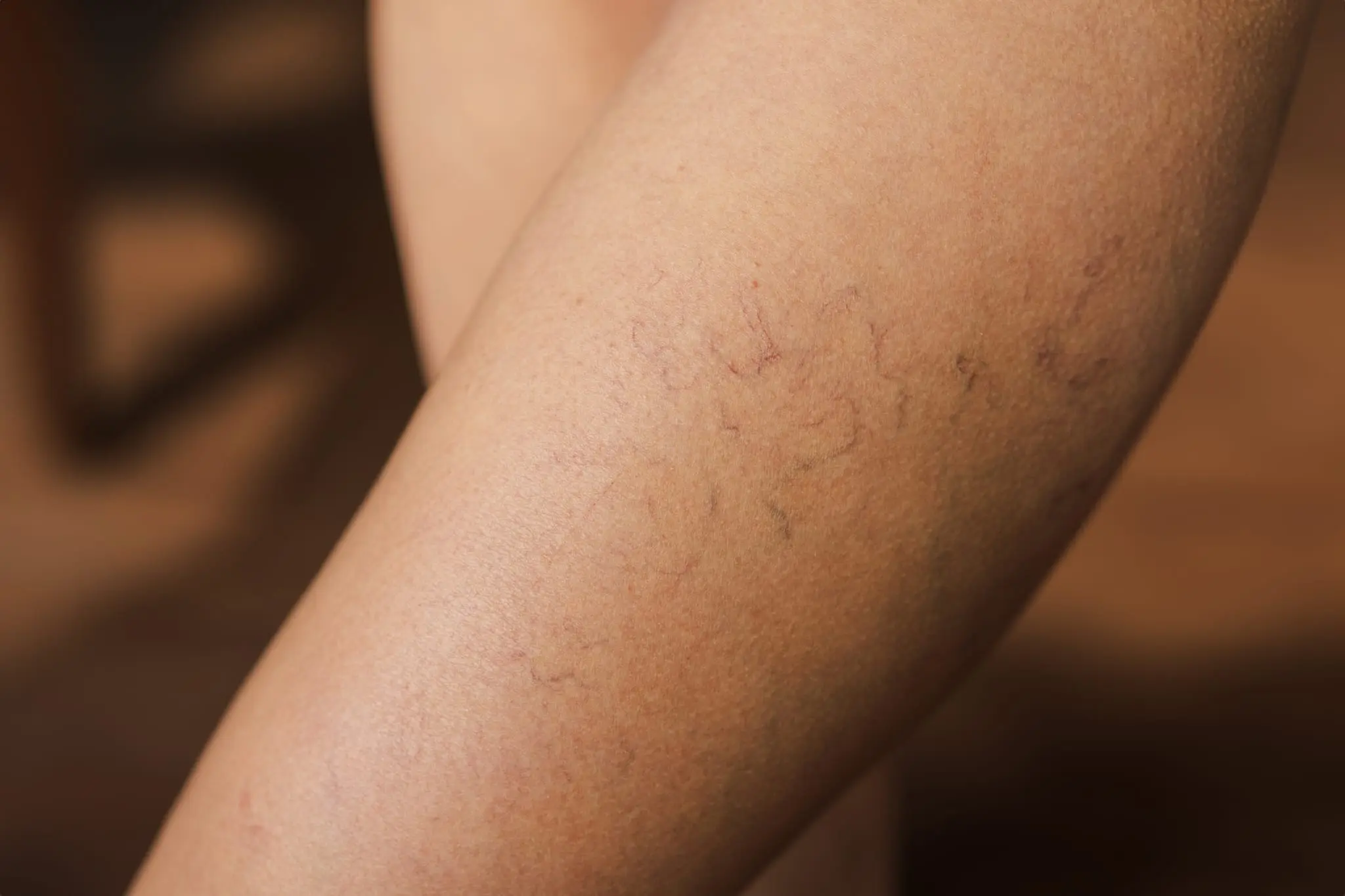
Varicose veins are the visible veins under the skin of the legs.
The veins in the legs are responsible for carrying blood from the feet and legs back to the heart. There are two types of veins:
- Deep Venous System
- Superficial Venous System (Great Saphenous Vein)
The deep venous system is the primary vein of the leg that runs through the leg muscles. It carries blood back to the heart through the pumping action created by muscle contractions.
The superficial venous system runs above the muscles, just beneath the skin, and also collects blood from the skin's capillaries. To move blood upward against gravity, it contains a series of one-way valves that open only in the upward direction. The deep and superficial veins join together at the groin level and continue as a single vein toward the heart. The valve mechanism in the superficial vein (great saphenous vein) starts at this junction and extends down to the ankle.
If the valve mechanism in the superficial vein, especially at the groin level, becomes dysfunctional, blood begins to pool inside the vein and its diameter increases (internal varicose vein). As the vein widens, the valves can no longer close properly, causing a vicious cycle of further dilation. As blood pooling increases, pressure within the vein rises and is transmitted to the tiny capillaries beneath the skin. As a result, these capillaries become visibly raised and discolored, appearing on the skin as varicose veins.
- Swelling, pain, and a feeling of heaviness in the legs, especially at the end of the day after prolonged standing.
- Nighttime leg cramps.
- Discoloration (blue-black) around the ankle, itching, skin thinning and shininess, and loss of hair below the knees.
- Blood clots may form within enlarged varicose veins, leading to pain and vein inflammation (thrombophlebitis)
- In advanced cases, difficult-to-heal wounds may develop around the ankle area (venous ulcers)
- Varicose veins are a vascular condition and should be treated by a cardiovascular surgeon. Do not begin treatment without first consulting a vascular specialist.
- Your doctor should thoroughly question you about the symptoms mentioned above. A family history of varicose veins should also be inquired about, as genetic predisposition is important.
- A physical examination of both legs—from the groin to the ankle—should be performed while standing and lying down.
- To evaluate your venous system, a Doppler ultrasound (a vein ultrasound of the leg) must be conducted. This test should be done while standing and with sufficient time and attention, as your treatment will be based on its findings.
If the Doppler ultrasound does not reveal internal varicose veins and there are no severely dilated veins, preventive measures should be taken.
- Standing for long periods or sitting with your legs hanging down can worsen symptoms. In such cases, wearing knee-high compression stockings can provide relief. Compression stockings come in many styles and colors—some are virtually indistinguishable from regular stockings when worn under clothing. There are also sheer models that resemble thin hosiery and do not require you to change your style of dress. Your doctor will advise you on the appropriate compression level and length of stocking you need. When purchasing, your ankle and leg measurements will be taken to ensure a custom fit.
- There is no cure for varicose veins through medication. However, there are symptomatic medications that can help relieve discomfort. These medications reduce fluid leakage from dilated veins, thereby helping to alleviate swelling, pain, and cramps.
- Regular exercise can reduce symptoms and slow the progression of varicose veins. Exercises that target the upper leg and lower abdominal muscles—such as Pilates and yoga—are especially beneficial. Walking and swimming activate muscles that pump blood through the deep veins, helping you return home at the end of the day with more refreshed legs.
- Elevating your legs, especially during times of discomfort, can provide relief. Lying down and resting your legs against a wall at an angle greater than 60 degrees for 5–10 minutes, 3–4 times a day, promotes venous drainage and has a therapeutic effect.
If you have small varicose veins that do not require surgery, injection of a foamy substance into the vein using very fine needles (sclerotherapy) can eliminate the veins. For even smaller veins, radiofrequency ablation may be used. In many cases, a combination of both treatments is necessary. These procedures not only improve cosmetic appearance but also relieve localized symptoms.
If your Doppler ultrasound shows significantly enlarged superficial veins (great saphenous vein) and severe valve insufficiency inside the vein (greater than grade 3 venous insufficiency), surgical treatment is necessary. If surgery is recommended, delaying the procedure can lead to the formation of new varicose veins and may result in a more complicated surgical intervention in the future.
The goal of surgery is to remove the varicose superficial vein from circulation and redirect all venous blood flow through the deep vein system. Once the high pressure in the superficial vein is eliminated, the pressure transmitted to the subcutaneous veins decreases, preventing the formation of new varicose veins.
In classical varicose vein surgery, an 8–10 cm incision is made in the groin area to access the point where the deep and superficial veins meet. The superficial vein is separated from the deep vein, and a wire is inserted into it to strip the vein from the groin down to the knee. Varicose veins in the leg are usually removed through small incisions that often do not require stitches. After surgery, the leg is bandaged, and the patient typically stays in the hospital for one night. About one week of rest is generally recommended for recovery.
In minimally invasive varicose vein surgery, a sheath (cannula) is inserted into the superficial vein at the knee level under Doppler ultrasound guidance. A wire (catheter) is then advanced through this sheath into the vein. The position of the wire is confirmed by Doppler to ensure it is at the junction between the deep and superficial veins. Along the course of the vein, a saline solution is injected through tiny needles around the vein to create a protective fluid cushion. Radiofrequency energy is then delivered through the wire to close the vein from the inside. The surrounding fluid prevents heat from damaging adjacent tissues. The procedure ends with the removal of the sheath from the vein. There are no surgical incisions or stitches involved in this procedure.
Varicose veins are a treatable condition and should be managed by a specialist in cardiovascular surgery.